
Skin Histology

Description: Skin histology, including cells of the skin, thin skin vs thick skin, and keratinized vs non-keratinized epithelium.
Skin Histology
I. Overview of Integumentary System
§ Integumentary system consists of the skin and associated appendages: sweat glands, sebaceous glands, mammary glands, hair, and nails
§ Skin is the largest organ in the body and serves multiple functions
○ Protective barrier against physical trauma, chemicals, and UV radiation
○ First area round of immune defense against infectious organisms
○ Body temperature regulation
○ Sensing the outside world (tactile, thermal, pain)
○ Production of vitamin D
§ Skin consists of the following layers
○ Epidermis
○ Dermis

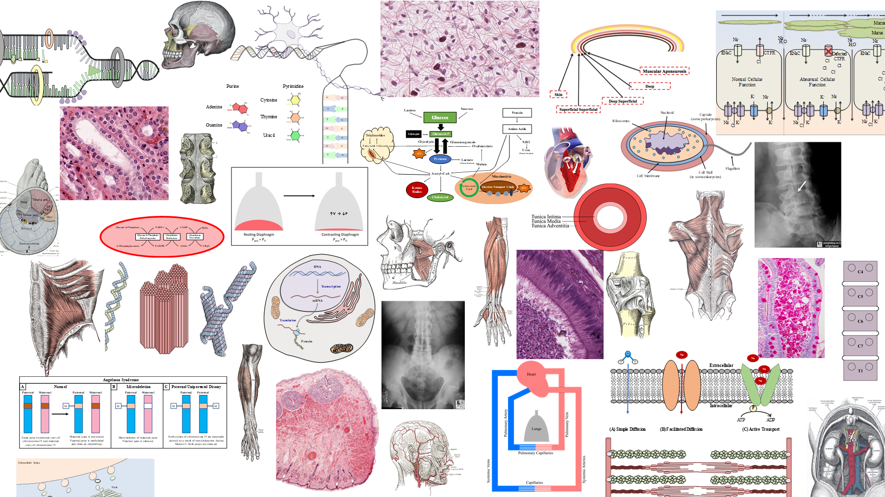
II. Epidermis
§ Most superficial layer of the skin that consists primarily of stratified squamous epithelium
○ Predominant cell is the keratinocyte
§ Consists of multiple layers:
○ Stratum basale (germinativum)
○ Stratum spinosum
○ Stratum granulosum
○ Stratum lucidum
○ Stratum corneum

III. Stratum Basale
§ Single layer of cuboidal epithelial cells at the base of the epidermal epithelium
○ Derived from the ectoderm
§ Contains germinal stem cells that continuously undergo mitosis to produce new keratinocytes
§ Also functions to connect the epidermis to the dermis via hemidesmosome connecting with integrins (basal lamina)
§ Histological appearance
○ Large oval nuclei
○ Basophilic cytoplasm


IV. Stratum Spinosum
§ Intermediate layer containing keratinocytes with a spiny appearance
§ Radiating bundles of tonofilaments (cytokeratin) forming desmosomes between cells
○ Spaces observed between cells are shrinkage artifact
○ Cells closer to the surface are flatter than cells closer to the stratum basale

V. Stratum Granulosum
§ Composed of keratinocytes that produce the stratum corneum layer
§ Basophilic inclusion bodies that contain keratohyalin granules composed of histidine- and cystine-rich proteins (filaggrin) that stimulate keratin filament aggregation
§ Undergo modified apoptosis, resulting in nuclear degradation, while maintaining cell structure

VI. Stratum Lucidum
§ Layer of thick stratum corneum composed of anucleate keratinocytes
§ Only found in areas of body with thicker skin, such as palms and soles of feet
§ Appear translucent and homogenous due to intracellular aggregation of keratin
VII. Stratum Corneum
§ Most superficial layer
§ Composed of anucleate (cornified) keratinocytes filled with keratin filaments and lamellar bodies
○ Coated with glycolipids that serve to create the water barrier feature of skin
§ Thick skin has a thicker, dense stratum corneum
§ Thin skin has a thinner, soft stratum corneum

VIII. Cells of the Epidermis
§ Keratinocytes
§ Melanocytes
§ Langerhans cells
IX. Keratinocytes
§ Epithelial cell of the epidermis
§ Produce keratin filaments that form bundles called tonofilaments that contribute to desmosomes linking adjacent keratinocytes
§ Undergo the process of keratinization that transforms granular cells into cornified cells
○ Involves increased aggregation of keratin filaments to form soft keratin
§ Stratum spinosum layer produces lamellar bodies which contain mixture of lipids for coating the epidermis to form the epidermal-water barrier

X. Melanocyte
§ Responsible for producing melanin
§ Derived from neural crest
§ Found between keratinocytes of the stratum basale, stratum spinosum, and within hair follicles
§ Attached to the basal lamina via hemidesmosomes
○ Not attached to keratinocytes (no desmosomes)
§ Histological appearance
○ Large ovoid nuclei
○ Pale staining cytoplasm

§ Produce melanin via oxidation of tyrosine to 3,4-dihydroxyphenylalanine (DOPA) to melanin
○ Melanin protects against damage from UV light
§ Multiple cytoplasmic extensions into the stratum spinosum
○ Facilitate transfer of filamentous melanin in melanosomes from melanocytes to keratinocytes via cytocrine secretion
§ Number of melanocyte cells is equal among all races
§ Skin and hair color are influenced by the degree of melanosome aggregation in keratinocytes
○ Rate melanin production, transfer of melanosomes, and lysosomal degradation vary among races
XI. Langerhans Cells
§ Dendritic macrophages that act as antigen presenting cells
○ Utilize major histocompatibility complex (MHC) II to present foreign antigens to T-cells
§ Able to travel to lymph nodes via dermal lymphatic vessels
§ Usually found in the stratum spinosum
§ Histological appearance
○ Dense, basophilic, indented nucleus
○ Pale cytoplasm
§ Electron microscopy reveals unique “paddle-shaped” intracellular granules known as Bierbeck bodies (unknown function)
XII. Merkel Cells
§ Cells found within the stratum basale layer that function as mechanoreceptors
○ Associated with unmyelinated nerve endings to provide touch sensation
§ Form attachments with adjacent keratinocytes via desmesomes
§ Histological appearance
○ Indented nucleus
○ Pale cytoplasm
○ Electron-dense, perinuclear vesicles
XIII. Dermis
§ Layer of skin deep to the epidermis with two layers:
○ Papillary dermis (superficial 1-20%) = loose connective tissue
○ Reticular dermis = dense irregular connective tissue
§ Composed of a fibrous network of collagen type I and III fibers with elastic fibers
§ Contains blood vessels, sweat glands, nerves, including sensory receptors
§ Site of immune response to infections, skin wounds, and cutaneous allergic reactions

I. Hypodermis
§ Underlying region composed of loose connective tissue and adipocytes
○ Provide cushioning and insulation
§ Some hair follicles and sweat glands may extend into the hypodermis
§ Sometimes contains smooth muscle

XV. Epidermal-Dermal Junction
§ Dermal papillae interdigitate with epidermal ridges to increase contact surface area between the two layers
○ Microanatomical basis for fingerprints and footprints
§ Most prominent in areas of skin that withstand significant shearing forces
○ Fingertips, palms, soles
XVI. Thick vs. Thin Skin
