
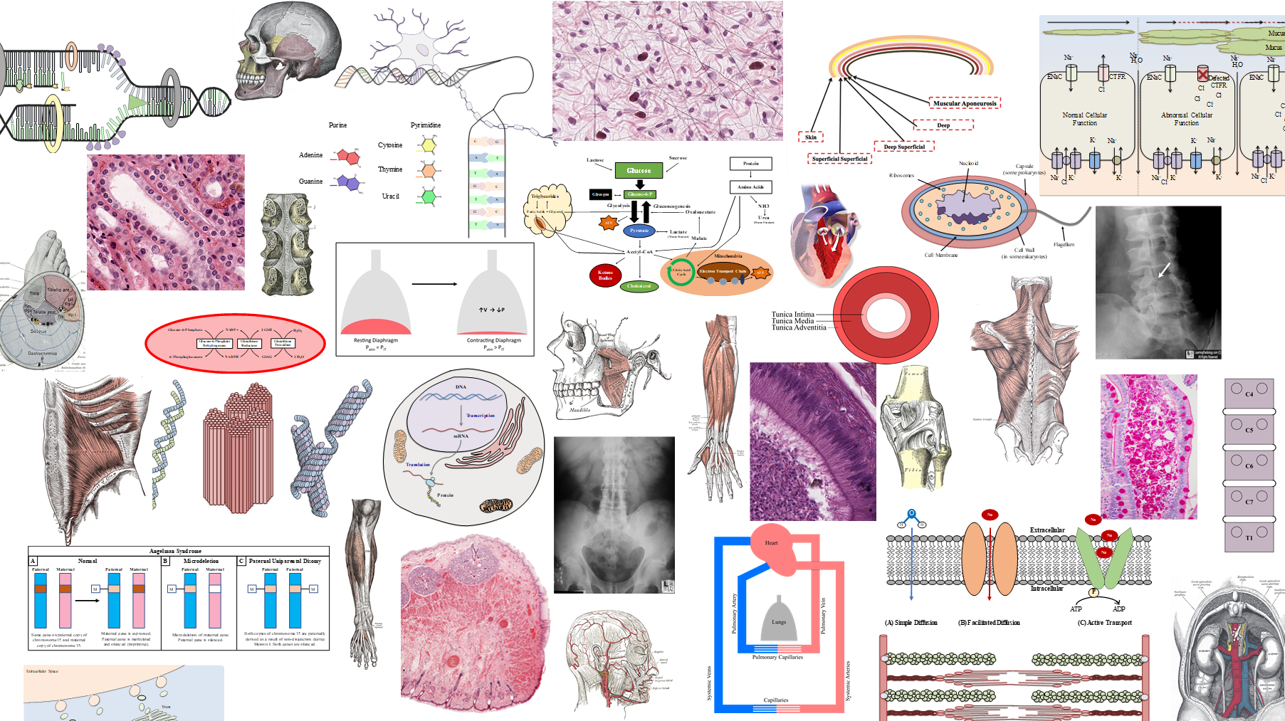
Microscopy & Staining
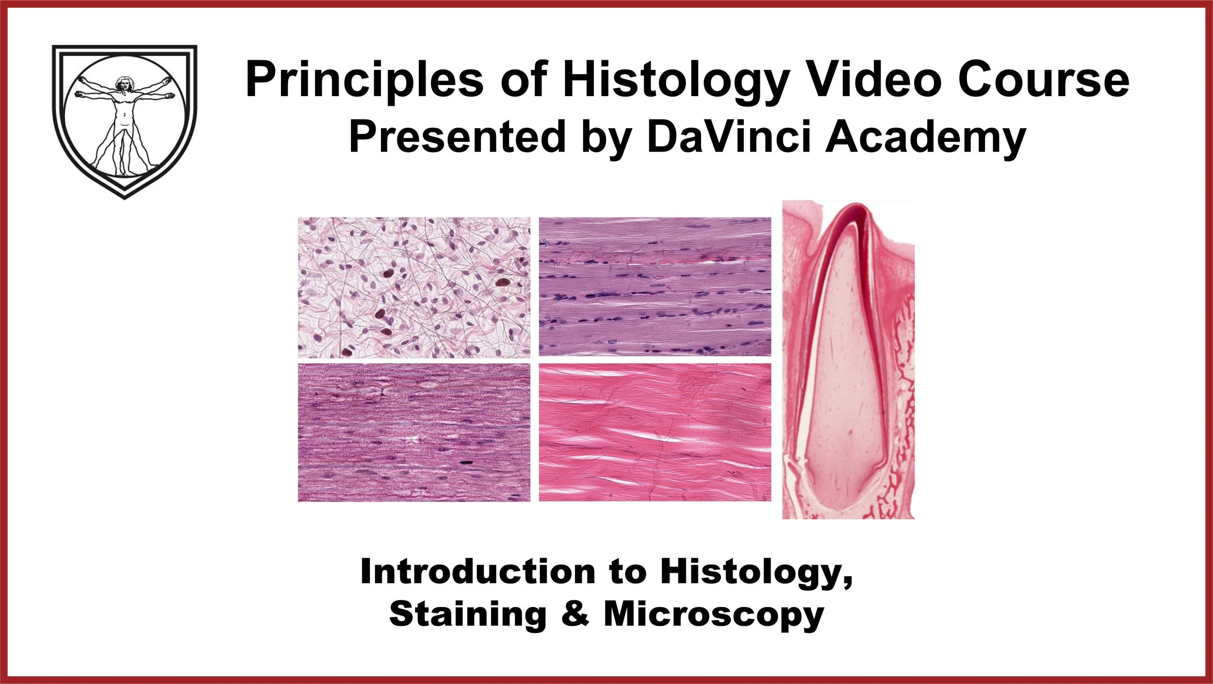
Description: Overview of histology, slide preparation, histological stains, and types of microscopy.
Microscopy & Staining
I. Histology
§ Study of Tissues
○ Tissues are made of cells and extracellular matrix (ECM)
§ Why study tissues?
○ We are made of tissues
○ To learn histology is to learn how we are made up and function
○ Injuries & pathologies occur at the atomic & molecular levels manifesting at the cell and tissue levels
○ At present, cell & tissue levels are most easily & economically accessible for diagnosis & to inform treatments
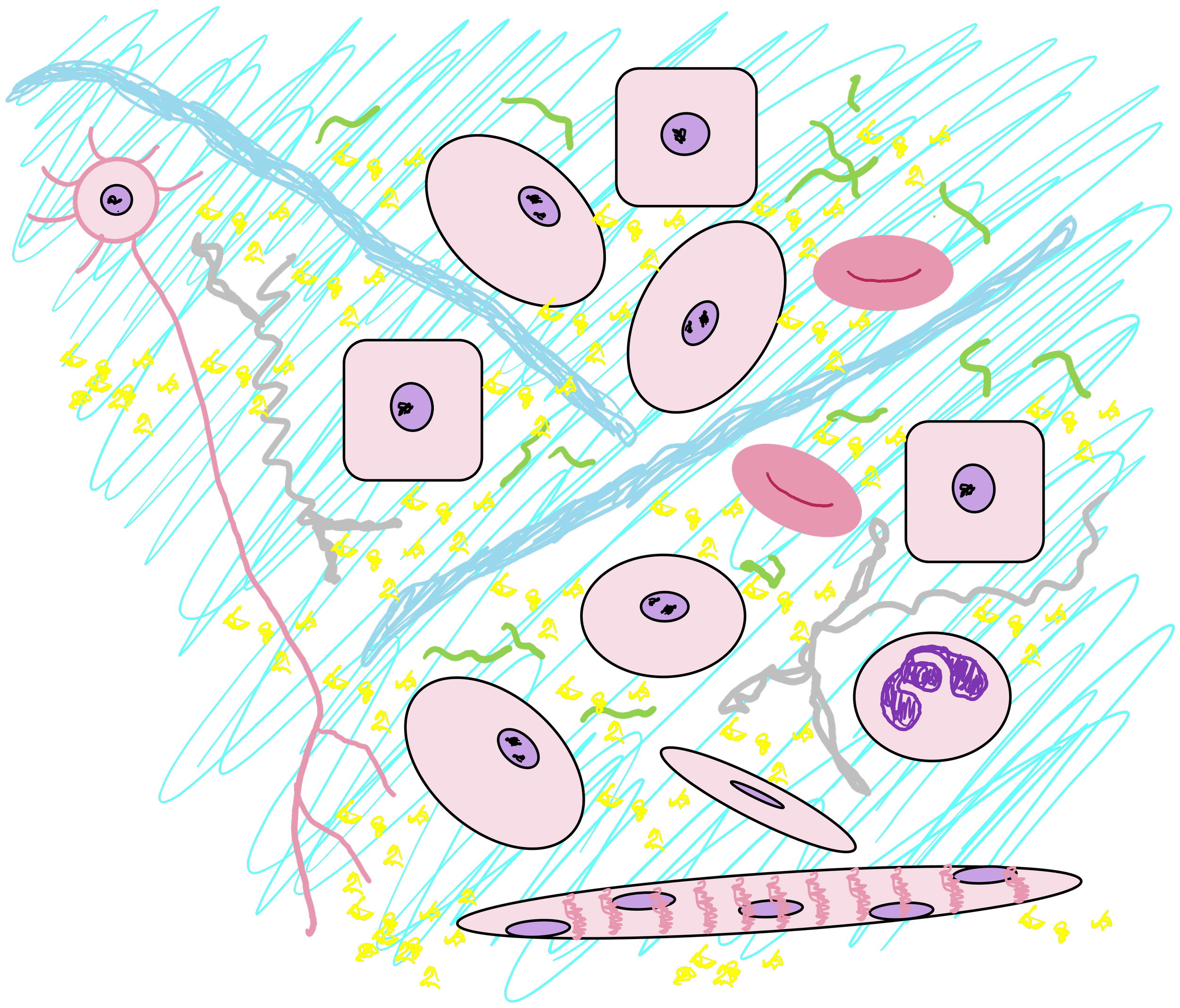
II. General Tissue Makeup
§ Cells
○ Variety of types
§ ECM
○ Fibers
‒ Collagen
‒ Elastic
‒ Reticular
○ Ground substance
‒ GAGs
‒ Glycoproteins
‒ Proteoglycans
○ Fluids
‒ Plasma-like, mostly water
§ If all tissues are made of the same “ingredients” why do various tissues look & function differently?
○ Types of cells
○ Proportion of ECM components
○ Different tissue types function together à Organ à Organ Systems à Anatomy
§ What technical steps are necessary to study tissues? Histotechnology
○ Tissue acquisition
‒ Cytological swabs
‒ Biopsies
‒ Surgical resections
○ Fixation
‒ Stops tissue degradation
‒ Most common fixative = formaldehyde (penetrates the tissue 1mm/1hr)
‒ Hardens & changes the color of the tissue
○ Tissue processing
‒ Replaces fat and water with hardening agent (paraffin)
○ Embedding
‒ Places the tissue in paraffin block
‒ Thoughtful embedding is important
○ Sectioning
‒ Paraffin block mounted on a microtome
‒ Each slice = 4-10 microns thick
○ Ribbon of thin paraffin slices is placed in water bath
○ Each section is mounted onto a glass slide
○ Staining tissue sections
‒ Remove paraffin
‒ Apply dyes and washing agents
‒ Place a glass coverslip on stained tissue
○ Tissue is ready for microscopic observation


III. Histotechnology: Dyes and Other Staining Agents
§ H & E: the most common combination of dyes
○ H = Hematoxylin
‒ Positively charged, basic dye à dyes negatively charged, acidic structures (DNA & RNA in nucleus & ribosomes)
‒ Has blue hue
‒ Structures that stain well are said to be basophilic for attracting basic hematoxylin
○ E = Eosin
‒ Negatively charged, acidic dye à dyes positively charged, basic structures (many proteins in cytoplasm)
‒ Has pink hue
‒ Structures that stain well are said to be eosinophilic or acidophic for attracting the acidic eosin
§ Histochemistry
○ Uses natural chemical interactions between the tissues and the chemicals
○ H&E is a type of histochemistry
○ Other histochemical stains:
‒ Trichrome: combination of dyes often used to visualize collagen
‒ Periodic Acid Schiff: reacts with carbohydrates to generate magenta color & is used to visualize carbohydrate-rich regions
Trichome = Tri (three) + chromo (color)
§ Immunohistochemistry
○ Uses specific interactions between antigen and antibody
○ Primary antibody applied to recognize the protein (antigen) of interest
○ Secondary antibody with a chromogen applied to recognize the primary antibody
○ Chemical applied to react with chromogen to turn the tagged region of tissue brown color
§ Immunofluorescence
○ Uses specific interactions between antigen and antibody, just like immunohistochemistry
○ Primary antibody applied to recognize the protein (antigen) of interest
○ Secondary antibody has fluorescent tag instead of a chromogen
○ Excitation by UV light or laser causes the fluorescent tag to emit specific frequency in a color spectrum
○ Can visualize more than one protein of interest


IV. Microscopy
§ Microscopes allow observation of prepared tissue slides at various levels of magnification
§ Light microscopy
○ Natural light illuminates the tissues for visual inspection
○ Used to observe tissues stained with histochemistry and immunohistochemistry
§ Phase contrast microscopy
○ Natural light illuminates the tissues for visual inspection
○ Leverages different refractory properties of cellular structures
○ Cells or tissues need not be stained à allows observation of live cells and tissues
§ Fluorescence microscopy
○ Works similar to light microscopes but has additional laser or UV light source
○ Used to observe tissues stained with immunofluorescence
§ Transmission Electron Microscopy (TEM)
○ Instead of photons, electrons are used as a source of “illumination”
○ Small size of electrons allow greater magnification and resolution of tissues and cells
○ Electrons pass through the tissue, casting different shades of “shadows”
‒ Darker areas on TEM (electron dense areas): more cellular or tissue materials are concentrated
‒ Lighter areas on TEM (electron light areas): less cellular or tissue materials are present & allowed electrons to pass
§ Scanning Electron Microscopy (SEM)
○ Instead of electrons passing through the tissue, they are scattered by the tissue
○ Able to capture three-dimensional surface contours of the tissue
§ Virtual Microscopy
○ Software or app simulating the use of a microscope on a digital screen
○ Allows navigation of the virtual tissue slide similar to Google Map
V. Clinical Application of Histotechnology
§ Critical component of pathology practice
○ Biopsies or surgical resections are documented & gross observations noted
○ Large surgical specimens are dissected, photographed and placed in a fixative for appropriate amount of time
○ Fixed specimens are sampled and sampled tissues are processed, embedded, sliced, mounted and stained
○ Diagnosis is made by a pathologist upon visual inspection of the tissues
○ Additional tissue processing, such as immunohistochemistry or immunofluorescence is ordered for ambiguous tissues or to confirm diagnosis
§ During certain surgical procedures, a surgeon may request a rapid tissue observation by a pathologist to ensure that surgical margin is free of malignancy or that a lymph node is free of metastatic cancer cells
○ The tissue is frozen rapidly and sectioned using a cryostat then stained
○ The quality of stained tissue is not as good as routinely processed tissue but can be an effective reference when speed is of importance
○ The frozen tissue is processed for paraffin section for confirmation at a later time
§ Critical component of pathology practice
○ Biopsies, surgical resections, and peripheral blood smears require a pathologist to review a histopathological slide from the tissue collected to make a diagnosis
○ Additional tissue processing, such as immunohistochemistry or immunofluorescence is ordered for ambiguous tissues or to confirm diagnosis
§ During certain surgical procedures, a surgeon may request a rapid tissue observation by a pathologist to ensure that surgical margin is free of malignancy or that a lymph node is free of metastatic cancer cells
○ The quality of stained tissue is not as good as routinely processed tissue but can be an effective reference when speed is of importance
○ Commonly used by surgical oncologists and dermatologists who specialize in Mohs surgery
VI. Histology Pro Tips
§ Magnification vs. Field of View
○ Low mag = more field of view but low on details
○ High mag = small field of view but high on details
○ Best practice: Start with low mag then incrementally go to high mag


§ Characteristic landmarks and unique architecture will easily give away its location

§ However, too much detail and too small a field of view can be disorienting
§ Histology requires context & perspective
§ Same types of tissues may appear differently
○ Colorful, sepia, grayscale, younger, older, low & high BMI; but recognizable as an Elvis
○ Even an impersonator can be identified

§ Artifacts are a natural part of histology
○ Many steps in histotechnology and staining, each step an opportunity to introduce artifacts
○ Tissue tearing, shrinkage, folding, fading stains, foreign particles
○ Maybe advantageous in tissue ID

§ Mind the representation
○ One, 7 micron slice of a large organ does not represent the entire organ

§ Typical tissue images are 2D representation of 3D structures
○ Levels of section
○ Planes of section
§ Need to develop skills to extrapolate 3D shape from 2D images (Mental gymnastics)

Post-Lecture Quiz
1) Which staining technique is best suited to visualize two proteins of interest on the same tissue section?
(A) H&E
(B) Immunofluorescence
(C) Immunohistochemistry
(D) Periodic acid Schiff
-
(A) Circle #1 encircles the nucleus of a cell
(B) Circle #2 encircles the nucleus of a cell
(C) Circle #3 encircles the entire cell

§ As one becomes a histology pro…
○ Able to recognize more characteristic architectures from higher mag / small field images
○ Able to use situational or surrounding clues
○ Acquire discerning eye for different versions and even impersonators

○ Able to recognize more characteristic architectures from higher mag / small field images
○ Able to use situational or surrounding clues
○ Acquires discerning eye for different versions and even impersonators
○ With tissue slides

§ To be a pro, one must visit as many sites (tissues) as many times as possible
○ Look at them from different angles (plane of sections) and in different lights (staining & microscopy)
Pre-Lecture Quiz
1) What is the subject of study in histology?
(A) Cells
(B) Diseases
(C) Organs
(D) Tissues
-
(A) Cells – Cytology is the study of cells
(B) Diseases – Pathology is the study of diseases
(C) Organs – Study of organs is at the anatomic level
(D) Tissues – Histology is the study of tissues

3) Which circle in the image outlines a cell?
(A) Circle #1
(B) Circle #2
(C) Circle #3
-
(A) Circle #1 encircles the nucleus of a cell
(B) Circle #2 encircles the nucleus of a cell
(C) Circle #3 encircles the entire cell
5) Which microscopy has the highest magnification power?
(A) Light microscopy
(B) Phase contrast microscopy
(C) Fluorescence microscopy
(D) Transmission electron microscopy
(E) Scanning electron microscopy
-
(A) Light microscopy - up to 1000x magnification
(B) Phase contrast microscopy – magnification power is typically less than A
(C) Fluorescence microscopy – similar magnification power as light
(D) Transmission electron microscopy – provides the highest magnification (1,000,000x)
(E) Scanning electron microscopy – up to 30,000x magnification
-
(A) H&E – stain acidic & basic structures in the cell indiscriminately
(B) Immunofluorescence – using secondary antibodies with different fluorescence tags, more than one protein may be tagged in a single tissue slide
(C) Immunohistochemistry – the chromogen on the secondary antibodies turn one color (brown) thus one can’t distinguish different protein expressions in a single tissue slide
(D) Periodic acid Schiff – this turns all carbohydrate-rich regions of the tissue magenta indiscriminately

3) Which circle in the image outlines a cell?
(A) Circle #1
(B) Circle #2
(C) Circle #3
5) Which staining technique is best suited to visualize two proteins of interest on the same tissue section?
(A) H&E (Hematoxylin & Eosin)
(B) Immunofluorescence
(C) Immunohistochemistry
(D) Periodic acid Schiff
-
(A) H&E – stain acidic & basic structures in the cell indiscriminately
(B) Immunofluorescence – using secondary antibodies with different fluorescence tags, more than one protein may be tagged in a single tissue slide
(C) Immunohistochemistry – the chromogen on the secondary antibodies turn one color (brown) thus one can’t distinguish different protein expressions in a single tissue slide
(D) Periodic acid Schiff – this turns all carbohydrate-rich regions of the tissue magenta indiscriminately
7) Which step in histotechnology stops tissues from degradation?
(A) Embedding
(B) Fixation
(C) Processing
(D) Sectioning
(E) Staining
-
(A) Embedding – processed tissue is placed in a paraffin block
(B) Fixation – fixative such as formaldehyde denatures proteins and stops tissue degradation
(C) Processing – fat and water in the tissue are replaced with paraffin
(D) Sectioning – tissues in paraffin block are thinly sectioned
(E) Staining – thinly sectioned tissues are stained

2) What is the most commonly used staining technique in histology?
(A) H&E (Hematoxylin & Eosin)
(B) Immunofluorescence
(C) Immunohistochemistry
(D) Periodic acid Schiff
-
(A) H&E (Hematoxylin & Eosin) is the most commonly used staining
(B) Immunofluorescence is used to visualize specific protein expression using fluorescent tags
(C) Immunohistochemistry is similar to immunofluorescence but uses chromogenic tags
(D) Periodic acid Schiff is used to visualize areas of high carbohydrate concentration
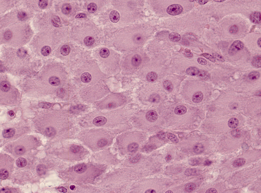
4) Which dye is responsible for the pink appearance of the cells in this image?
(A) Eosin
(B) Hematoxylin
(C) Periodic Acid Schiff
(D) Trichrome
-
(A) Eosin, a negatively charged acidic dye, stains positively charged proteins in the cytoplasm
(B) Hematoxylin – a positively charged basic dye stains negatively charged acid molecules
(C) Periodic acid Schiff reacts with carbohydrate moieties
(D) Trichrome stains collagen fibers

2) Which circle in the image outlines a cell?
(A) Circle #1
(B) Circle #2
(C) Circle #3
-
(A) Circle #1 is a nucleus of a cell, the DNA (deoxyribonucleic acid) in the nucleus stain well with hematoxylin which has blue to purple hue
(B) Circle #2 is the cytoplasm of a large cell and is staining pink with eosin which stain positively charged, basic substances
(C) Circle #3 is an artifact
*The pale staining areas are rich with ground substance
4) To compare the amount of glycogen storage in the liver cells of starved vs. well-fed rats, which staining technique should be used?
(A) H&E (Hematoxylin & Eosin)
(B) Immunofluorescence
(C) Immunohistochemistry
(D) Periodic acid Schiff
-
(A) H&E – stain acidic & basic structures in the cell indiscriminately
(B) Immunofluorescence – using secondary antibodies with different fluorescence tags, more than one protein may be tagged in a single tissue slide
(C) Immunohistochemistry – the chromogen on the secondary antibodies turn one color (brown) thus one can’t distinguish different protein expressions in a single tissue slide
(D) Periodic acid Schiff – this turns all carbohydrate-rich regions of the tissue magenta indiscriminately
6) Which structure is a form of ground substance?
(A) Collagen
(B) Glycosaminoglycans
(C) Reticular fibers
(D) Plasma
-
(A) Collagen - a type of fiber
(B) Glycosaminoglycans – one form of ground substance, the others are proteoglacans, and glycoproteins
(C) Reticular fibers are a type of fibers
(D) Plasma is the fluid component of blood which is similar to the interstitial fluid